By
Ayse Guner
Arizona Daily Wildcat
UMC's mini-medical school teaches about the body, organs and diseases
"Lub. Lub dub. Lub. Lub dub."
The closure of the atrioventricular valve - the heart's key valve that makes a "lub" sound every time the blood travels from the left to the right chamber - is what doctors listen to with a stethoscope. Then the second sound comes in, the "dub," with the closure of the pulmonary valve, another key valve in the heart.
This blood-flow system in the heart, which allows a person to live, works just like a water balloon. When the pressure condenses the heart from the middle, valves keep blood flowing in the right direction so the blood can reach the destined organs to transport water, oxygen, salts, proteins and blood cells.
If a person has a bad valve, doctors hear a different sound, one like "lavusha, lavusha," said Christopher Leadem, associate professor of cell biology and anatomy at UMC.
The University Medical Center, with instructors such as Leadem, provides "mini-medical school" classes in an attempt to teach the public about the body and its organs. Attendees dissected a pig heart two weeks ago, learned about heart disease last week and will discover the operations of the brain and nervous system tomorrow night.
Although a pig heart is almost twice as large as a human heart, the structures are the same, Leadem said as he invited the course attendees to an anatomy lab on the third floor of UMC after a one-hour lecture on the organ.
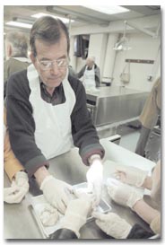
Leadem handed clean, white aprons to his students as he walked through the lab, leading them to tables where about 30 limp pig hearts lay.
Soon, the students would dissect these hearts, which cost about $3.50 each, into bits and pieces.
Local physical therapist Virginia Morgan, decked out in her apron, anxiously organized her group of four. Morgan was the most experienced one in her group, having dissected a human heart about 30 years ago.
"But this is huge," she said, holding the pig heart in one hand and a knife in the other.
Across each table, a monitor hung from the ceiling, displaying a highly-magnified landscape of the heart Leadem was dissecting.
During the lecture, Leadem stressed that slides of the heart would not tell much about how the organ functions.
"We have to dissect it. It doesn't tell," he said.
"We need to take things apart," Leadem added. "We can't have anatomy without taking things apart."
With this in mind, Morgan slide her knife into the top part of the organ, cutting the fatty parts first, then working her way into the vessels and reaching down into the pulmonary artery.
"Feel that atrium," she said to her colleague at the table, Jody Hugher, who hunched over the organ with wide eyes.
"How much blood would enter this?" Hugher asked Morgan.
"All the blood," Morgan answered. "The entire body's blood goes through this heart."
Morgan probed her finger through one side of the heart's entry to another. She illustrated how the valves worked and how fibers connected parts of the organ.
"This is solid. There is no hole in it," Morgan said about the valve she was touching.
Meanwhile, the screen of the monitor reflected the inside of Leadem's dissected organ. He pulled the heart apart, explaining the role of the arteries, where blood passes through and how the valves shut, as well as how they prevent the blood flow back into the restricted areas.
The teams worked quickly, even though some had never studied anatomy. Varying questions were raised, such as where the pig hearts came from, how much each was worth and where the human cadavers were stored.
Leadem would not allow the "mini-med school" students to look at highly-dissected human cadavers stowed in the back of the room.
Those were for UMC's medical students, Leadem said, adding that they use about 30 donated human bodies in one anatomy class, which is offered only one semester a year.
"Pig hearts are more available than human," Leadem said. "You can order them easier."
Curiosity in cadavers and anatomy has led to major medical discoveries in past centuries, leading up to today's era of bypass surgery and transplanted hearts, Leadem said.
To understand the sounds it makes, its functions and malfunctions, the heart has been dissected for the past 2,200 years.
During the Renaissance period, artists such as Leonardo Da Vinci and Michelangelo dissected entire human bodies to enhance the realism of their human drawings.
The first study of anatomy, though, began much earlier.
In 300 B.C., in the city of Alexandria, Egypt, two physicians named Herophilus and Erasistratus dissected about 800 bodies to study the structure of the human body, Leadem said.
"But the dark side of it was they dissected on living," he added.